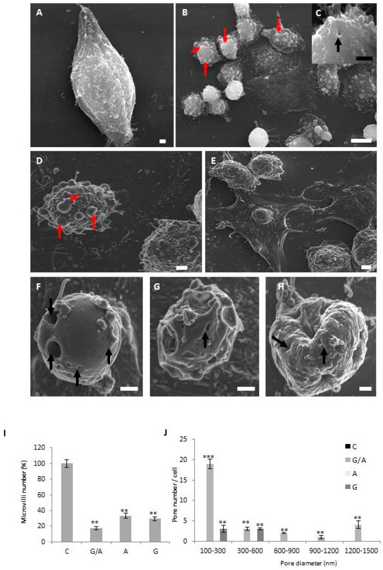
Figure 3.
Scanning electron microscopic images of parental and transduced DLD-1 cells. DLD-1 cell line adhered to the culture surface with multiple filipodium (A). DLD1/Tet-On-gef cell line showed cells with several apoptotic bodies (red arrows) (B) and some cells have pores on their membrane surface (C). DLD1/Tet-On-apoptin cells showed signs of apoptosis with cytoplasmic membrane disruption, many apoptotic bodies, and clear signs of substrate detachment (D). DLD1/Tet-On-gef-apoptin cells, like the others, produced dead cells without filipodium and abundant apoptotic cells with the surface covered with apoptotic bodies (E). The most curious thing is that many more cells appeared with the surface full of pores of different sizes (F–H). Red arrows signal apoptotic bodies and black arrows pores (Scale bar A, C, D, F, G, H = 1 μm; Scale bar B = 10 μm; Scale bar E = 2 μm). Quantification of filipodium number (I). Pore number (J). The height of each bar represents the number of pores with diameters in the interval between its lower and upper bounds on the x-axis. Pore diameters between 100–1500 nm can be observed in DLD21/Tet-On-gef-apoptin cells. Quantifications were measured from the SEM images (5 random photos) from three independent sample preparations. The data is represented as mean ± S:D (** p < 0.01 vs. control and *** p < 0.001 vs. control).

