FIGURE 3.

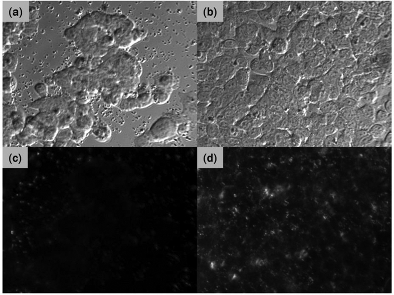
Brightfield images of (a) EGFR-negative (H520) cells and (b) EGFR-positive (A549) cells. (c) and (d) show the corresponding NIR darkfield images of cells incubated with UCNPAuNR nanoclusters and then washed off with PBS.

Brightfield images of (a) EGFR-negative (H520) cells and (b) EGFR-positive (A549) cells. (c) and (d) show the corresponding NIR darkfield images of cells incubated with UCNPAuNR nanoclusters and then washed off with PBS.