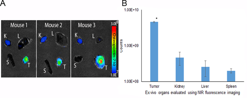
Figure 6.

Secondary confirmation ex vivo using near infrared fluorescent imaging. Imaging demonstrated ex vivo increased probe accumulation within the breast tumor compared to the kidney, liver and spleen at 3 h. A) NIR-fluorescence evaluation of tumor, kidney, liver and spleen using the AMI system indicated that osteopontin-750 accumulated selectively within the breast tumor. B) ROI measurements on organs ex vivo were grouped by organ among three mice. Osteopontin-750 accumulation within the tumor was 4.6 × 10⁹ (p = 0.0001) compared to the kidney (4.7 × 10⁸), liver (2.5 × 10⁸) and spleen (2.0 × 10⁸).
