Figure 1.
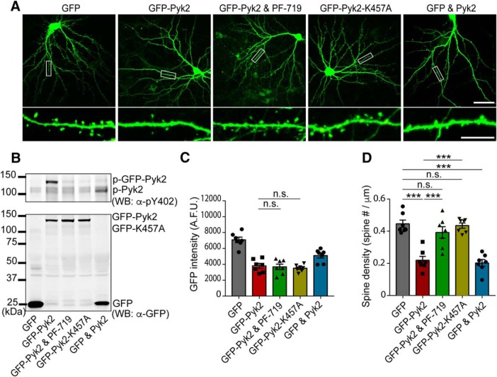
Pyk2 induces dendritic spine loss. A, Representative GFP fluorescent images of cultured mouse hippocampal neurons. Neurons were transfected with GFP alone, GFP-Pyk2, GFP-Pyk2 with 1 μm PF-719, GFP-K457A, or GFP and Pyk2 (untagged) at DIV 14 and then fixed at 21 DIV. Bottom, Enlarged images of the enclosed rectangles on the top. Scale bars: low-magnification (top), 50 μm; high-magnification (bottom), 10 μm. B, Lysates from transfected neurons were subjected to Western blotting with anti-GFP and anti-p-Pyk2 Y402 antibodies. C, GFP intensity quantification from imaged neurons for spine quantitation in A. D, Quantification of dendritic spine density in the transfected neurons. Data are presented as mean ± SEM (GFP, n = 10; GFP-Pyk2, n = 11; GFP-Pyk2 and PF-719, n = 9; GFP-K457A, n = 10 coverslips from 3 different cultures). ***p < 0.001 by one-way ANOVA, Tukey's multiple-comparisons test.

