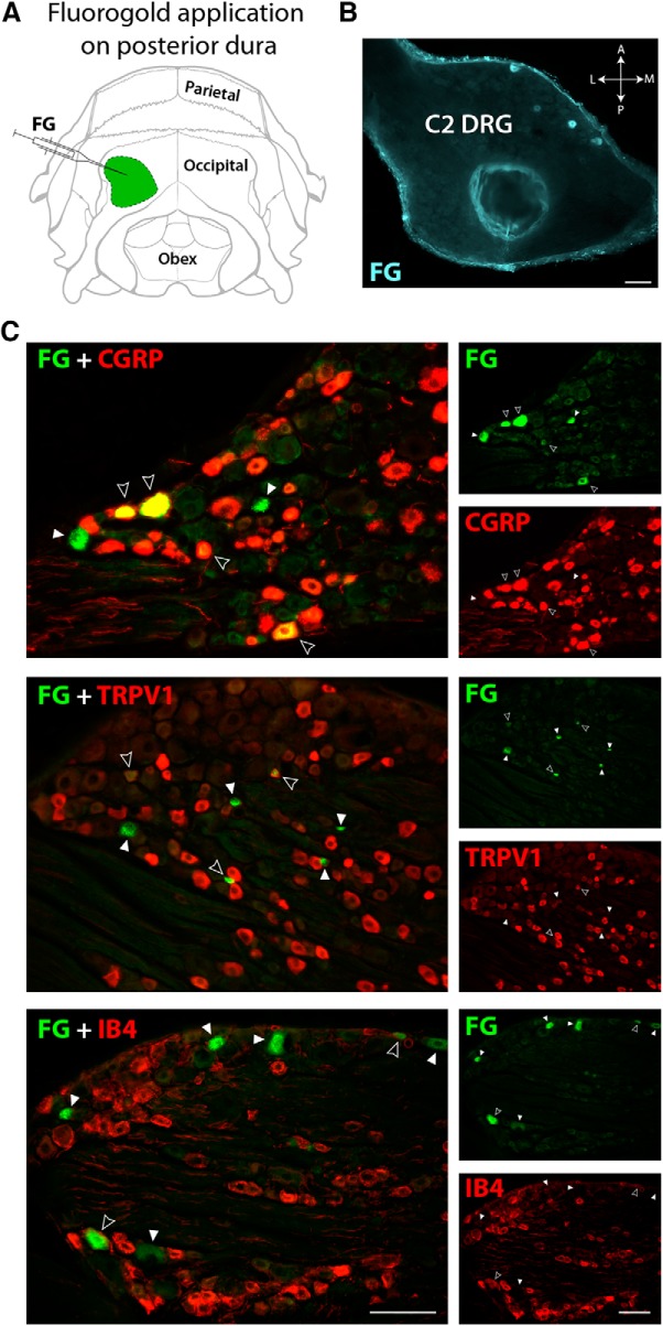
Figure 3.
Retrograde labeling in C2 DRG from the posterior dura. A, Illustration of the rat's skull view from behind showing the area of FG application (green) on the posterior dura. B, Transversal view of a C2 DRG section showing retrogradely-labeled neurons filled with FG near the anterior edge of the left ganglion. C, A proportion of FG-labeled neurons (green) in C2 DRG was also immunoreactive to CGRP, TRPV1, or IB4 (red). Images in the left column were created by superimposition of the images in the right column. Filled arrowheads point to FG-positive cells. Open arrowheads indicate double-labeled cells with FG and with each of the three markers of sensory neurons used (yellow). Scale bars, 100 μm.

