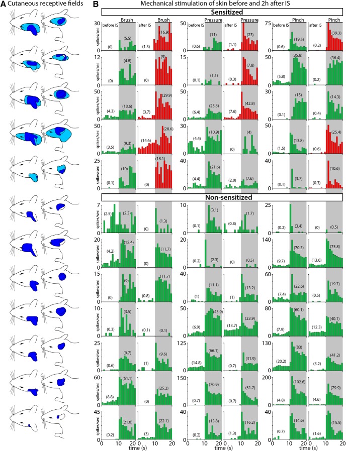
Figure 8.
RFs and neuronal responses to innocuous and noxious mechanical stimulation of the skin. A, Illustration of the rat's head and neck showing the cutaneous RFs of C2–C4 spinal cord dura-sensitive neurons measured during mechanical stimulation at baseline (dark blue) and 2 h after application of IS (light blue). B, Responses of individual neurons to mechanical stimulation (brush, pressure, and pinch) of the skin before (left columns) and after IS (right columns). Neurons were classified as sensitized (red) if their response magnitude increased by at least 50% after IS. Numbers in parenthesis represent firing rate in mean spikes/s during baseline (10 s) and stimulation (10 s). Shaded areas indicate the period of mechanical stimulation.

