
Fig. 3.
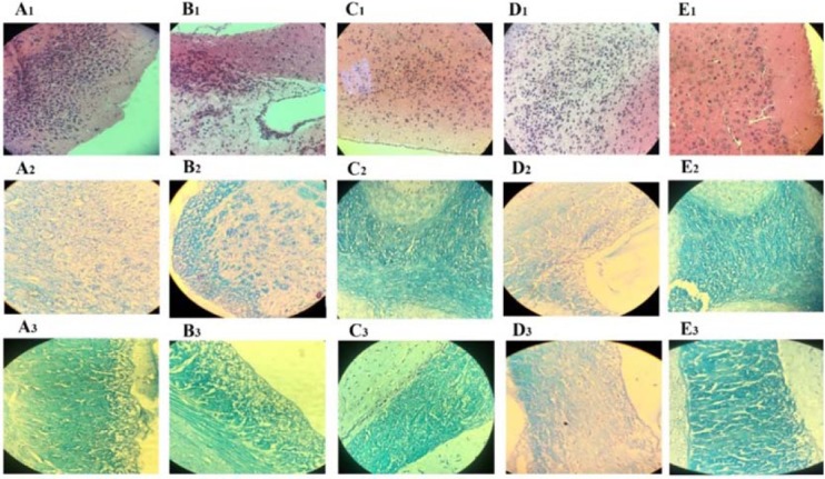
Histological findings of brain sections of mice. Group A, RAPA + HSO/EPO-treated mice show (A1) extensive infiltration of inflammatory cell, neurophagia, (A2) spongy tissue, and (A3) extensive demyelination; group B, RAPA-treated mice show (B1) focal infiltration of inflammatory cells, (B2) spongiotic zones, and (B3) demyelination; group C, HSO/EPO-treated mice show (C1) a few inflammatory cells (C2,3) without spongy lesions and demyelination; group D, control EAE mice show (D1) a great number of inflammatory cells, (D2) extensive degeneration, spongy tissue, and (D3) demyelination; group E, the section of the brain of naive mice exhibiting (E1-3) no clinical signs. The first row was stained with H&E and the second and third rows were stained with LFB. EAE, experimental autoimmune encephalomyelitis; HSO/EPO, hemp seed oil/evening primrose oil; RAPA, rapamycin; H&E, hematoxylin and eosin; LFB, luxol fast blue.