
Figure 4.
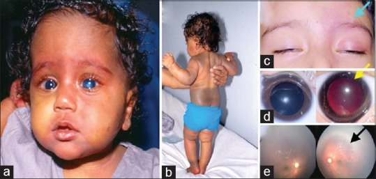
(a) Clinical photograph of a child with bilateral congenital glaucoma, bilateral vascular nevus on the face, and (b) extensive pigmentary nevus on the skin (Phacomatosis pigmentovascularis). (c) External photograph of a child with unilateral Sturge–Weber syndrome (Left-sided facial hemangioma). (d) Right and left eye photographs showing a dark red glow in the left eye suggestive of choroidal hemangioma. (e) Fundus photograph showing diffuse choroidal hemangioma in the left eye