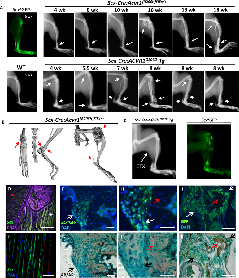
Fig 2. Expression of mutant ACVR1R206H or ACVR1Q207D alleles in Scx-lineage tendon-resident progenitor cells results in spontaneous peri-articular, ligament, and tendon ossification.

(A) Scleraxis expression is localized to tibiotalar ligament, patellar and Achilles tendons as observed by ex vivo fluorescence in Scx-GFP transgenic mice. Spontaneous ossification of tibiotalar ligament, patellar tendon and Achilles tendon progresses slowly in Scx-Cre:ACVR1R206H mice from 4 through 18 wks, with rapid progression in Scx-Cre:ACVR1Q207D-Tg mice from 4 through 8 weeks (B) Micro-CT of Scx-Cre:ACVR1R206H mice reveals distinct ossification of the tibiotalar ligament, Achilles tendon, and periarticular ossification, but no intramuscular ossification. (C-D) Scx-Cre:ACVR1Q207D-Tg:Rosa26-YFP mice injected with CTX (P21, gastrocnemius) develop typical tendon and ligament ossification, as seen by x-ray (P42) corresponding to areas of Scleraxis expression via Scx+YFP fluorescence but lack evidence of any intramuscular ossification (0/5 transgenic mice injected). (E) Cryosections of fibular head counterstained for CD45 (magenta) reveal Scleraxis expression (green) at ligamentous insertions (white arrow) but not in adjacent skeletal muscle (red arrow) in Scx-GFP transgenic mice. The HO lesions infiltrating the (F) talonavicular ligament, (G) talonavicular ligament in higher magnification, and (H) patellar tendon in Scx-Cre:ACVR1Q207D-Tg;Rosa26-YFP mice reflect the contribution of Scx+YFP+ cells to nearly all Alcian Blue (AB)-stained hypertrophic chondrocytes and heterotopic cartilage (white arrows) in these lesions, but essentially no contribution to osteocytes or mineralized matrix (red arrows) stained with Alizarin Red (AR), with DAPI as a nuclear counter-stain.
