
Fig 4. Mx1-lineage muscle interstitial but not bone marrow cells are sufficient for intramuscular HO.
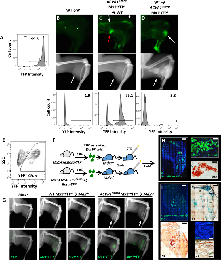
(A-D) Reciprocal bone marrow transplant experiments demonstrate medullary expression of mutant ACVR1 is dispensable for HO. (A) Control Mx1-Cre:Rosa26-YFP or mutant Mx1-Cre:ACVR1Q207D-Tg:Rosa26-YFP mice treated with pIpC exhibited >90% YFP labeling of marrow cells. Control or mutant mice were preconditioned antenatally with busulfan (E16, maternal i.p. injection), and engrafted at P2 with total bone marrow cells (5 ×10⁵ cells i.p.) harvested at P21 from WT or mutant donor mice previously treated with pIpC (P7-P21). In contrast to WT mice engrafted with WT marrow (B), WT mice engrafted with mutant marrow (C) exhibited a high percentage of YFP+ bone marrow comparable to mutant donor mice (A) by flow cytometry 78.2% ± 15.9% (n=5), and ex vivo fluorescence (white arrows), but did not exhibit injury-dependent ossification following CTX, despite the presence of infiltrating YFP+ cells due to CTX-induced inflammation (red arrow). Conversely, Mx1-Cre:ACVR1Q207D-Tg:Rosa26-YFP mice engrafted with WT marrow (D) exhibited very low frequencies of residual YFP+ bone marrow by flow cytometry (bottom panel, 0.5–5%; n=11), but exhibited robust CTX-induced intramuscular ossification with YFP+ lesions (white arrows, top and middle panel). (E-I) Mx1+ lineage muscle interstitial cell engraftment studies demonstrate Mx1+ lineage interstitial cells are sufficient for injury-dependent intramuscular HO. Mx1+YFP+ (5×10⁵) cells sorted from the muscles of P21 control Mx1-Cre:Rosa26-YFP or mutant Mx1-Cre:ACVR1Q207D-Tg:Rosa26-YFP mice previously treated with pIpC (P7-P19) were transplanted into gastrocnemius muscles of Dmdmdx−5cv:Rag1null (Mdx−/−) mice (P21) in Matrigel (E-F). In comparison to Mdx−/− control mice injected with Matrigel only (G, left), Mdx−/− mice injected with WT Mx1+YFP+ cells exhibited engraftment after 6 wks based on YFP fluorescence but no heterotopic ossification with or without injury (G, middle panel, no lesions seen in 5 treated mice), whereas Mdx−/− mice injected with mutant Mx1+YFP+ cells exhibited engraftment and developed intramuscular ossification following CTX treatment (G, right panel, lesions seen in 3/5 mice treated). (H) Histological analysis of mice injected with WT Mx1+YFP+ control cells demonstrated engraftment of YFP+ cells interspersed in gastrocnemius and popliteal fossa, all of which stain with Oil Red O (ORO). (I) Mice engrafted with ACVR1Q207D Mx1+YFP+ cells demonstrated engraftment of YFP+ cells throughout HO lesions of the gastrocnemius, with mineralization evident by Alizarin Red (AR), formation of ectopic cartilage demonstrated by Alcian Blue (AB), but were notable for the absence of YFP fluorescence in heterotopic marrow, shown by fluorescence and DAPI counter-staining (inset top panel), and the co-localization of YFP fluorescence with mineralized areas (AR, inset bottom panel).