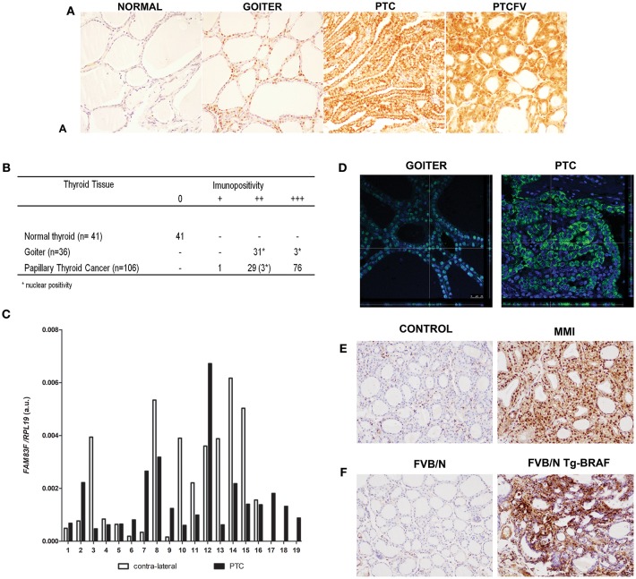
Figure 1.
FAM83F protein expression in human thyroid tumors and in animal models. (A) Detection of FAM83F using IHC shows strong cytoplasmic expression in papillary thyroid cancer (PTC), while in goiter strong nuclear expression is detected. Normal adjacent thyroid tissue is mostly negative for FAM83F staining. (B) IHC quantification in PTC (n = 106), goiter (n = 34), and normal thyroid tissue (n = 41). (C) FAM83F gene expression in a set of 19 paired human PTC vs. contra-lateral thyroid tissue by qPCR. Data shown as relative expression of FAM83F/RPL19. (D) Confocal microscopy of FAM83F in goiter and PTC. Immunofluorescence staining of FAM83F protein in goiter section (left panel) shows nuclear localization observed in the z-stack that colocalizes the nucleus with the green fluorescence of FAM83F, while in PTC section (right panel) FAM83F expression is cytoplasmic. (E) FAM83F protein expression in rat-induced pharmacological goiter. The treatment of rats with MMI for 5 days increased immunopositivity of FAM83F in thyroid follicular cells with strong positivity in the nucleus and mild positivity in cytoplasm (n = 5). (F) FAM83F protein expression in BRAFT1799A transgenic mice with PTC. Detection of FAM83F using IHC shows strong cytoplasmic and nuclear expression in 5–weeks old mice PTC (n = 5), while no expression is observed in normal thyroid (n = 4).

