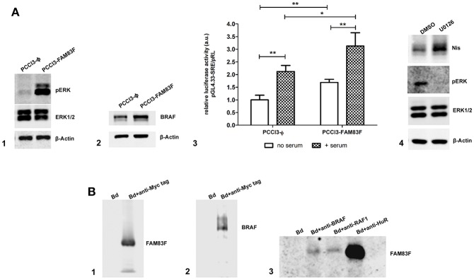
Figure 6.
FAM83F overexpression effect on MAPK signaling. (A) Analysis of MAPK signaling components protein expression and activation in rat thyroid follicular cells PCCL3 cells overexpressing FAM83F; (A1) Western-blotting of pERK and ERK1/2 levels; (A2) Western-blotting of BRAF protein levels; (A3) luciferase reporter assay with the pGL4.33[luc2P/SRE/Hygro] plasmid that contains serum-responsive elements activated by MAPK/ERK signaling in PCCL3-FAM83F after 2 h of serum addition to culture medium. Results are shown as mean normalized values (luminescence of pGL/ luminescence of pRL plasmid). *P < 0.05 and **P < 0.01. (A4) Western-blotting of Nis in PCCL3-FAM83F cells treated with MAPK signaling inhibitor U0126 at 5 uM for 48 h. (B) Immunoprecipitation (IP) in PCCL3-FAM83F cells using a anti-Myc Tag antibody to immunoprecipitate FAM83F protein (Myc-tagged); (B1) Detection of FAM83F protein levels in Myc-Tag IP lysate by WB; (B2) Detection of BRAF protein levels in Myc-Tag IP lysate by WB; (B3) Immunoprecipation of BRAF, RAF1, and HuR followed by detection of FAM83F in PCCL3-FAM83F cells by WB. Bd group stands for beads only IP (no antibody).

