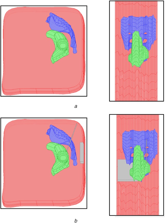
Fig. 2.

Four view of the three-dimensional finite element model created in COMSOL Multiphysics are shown
a Top row gives a sagittal (left) and axial (right) view of the model with the three segmented anatomical regions: Pelvis (pink), rectum (green) and sacrum (blue)
b Lower row shows the same views with the implanted electrode and IPG added to the model (grey)
