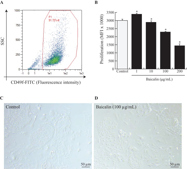
Figure 1. Effects of baicalin on cell proliferation in bovine MEC.
A prerequisite to this study was to validate our experimental model, notably by ensuring that isolated MEC corresponded to mature secretory cells. Hence, cells were dispensed at 500,000 and incubated in darkness at 4 °C for 30 min with Fluorescein isothiocyanate (FITC) anti-rat IgG1 CD49f (α6 integrin). CD49f positive cells are gated in red (A). Next, bovine MEC were incubated with different concentrations of baicalin (0, 1, 10, 100, 200 µg/mL) for 24 h in triplicate. (B) Cell proliferation was measured during 16 h using BrDU cell incorporation. MEC were plated in 96 well-plates at a density of 5,000/well. Increased Mean Fluorescence Intensity (MFI) values denoted higher proliferation rates in MFI (×1,000). Data shown as mean ± S.E. *p < 0.05 versus control (B) Phase contrast images of MEC in culture incubated with zero µg/mL of baicalin (C) and 100 µg/mL of baicalin (D). Black bar stands for 50 µm.

