
Figure 1. Human endothelial progenitor cells (EPC) produced extracellular mitochondria.
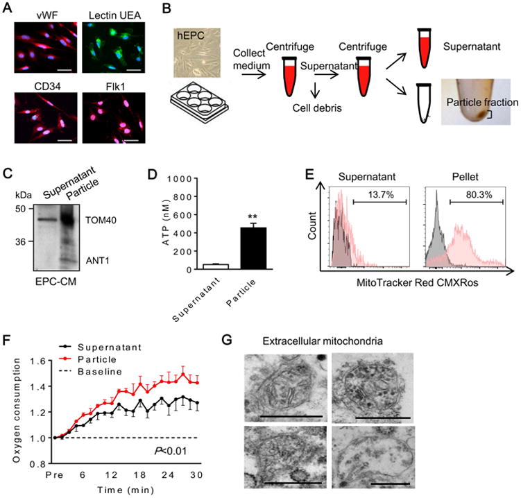
(A) Human EPC were identified by representative markers including vWF, lectin UEA, CD34 and Flk1. Scale: 50 μm. (B) Experimental design to separate fractions from EPC-derived conditioned medium. First centrifuge; 2,000 rpm for 10 min, second centrifuge; 12,000 rpm for 15 min. (C) Western blot confirmed mitochondrial proteins in EPC-derived extracellular fractions. TOM40; mitochondrial outer membrane protein, ANT1; mitochondrial inner membrane protein. (D) ATP measurement showed higher ATP content in pellet compared to supernatant (n=5). **P<0.01. (E) Flow cytometry analysis demonstrated that pellet highly contained MitoTracker positive particles compared to supernatant. (F) Oxygen consumption analysis in EPC-derived pellet and supernatant. EPC-derived pellet had higher oxygen consumption compared to supernatant (n=3). (G) Electron microscopy revealed that EPC-derived pellet contained mitochondria. Scale: 500 nm.