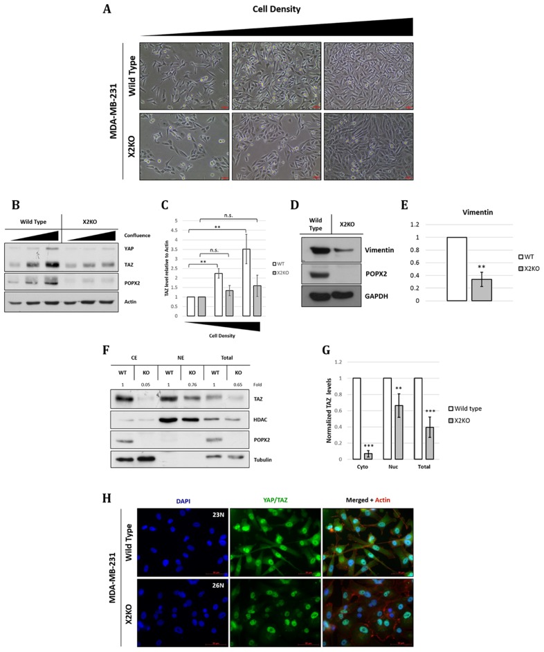
Figure 4. Loss of POPX2 affects MDA-MB-231 morphology a well as TAZ and vimentin protein levels.
(A) Brightfield images of MDA-MB-231 wild type and X2KO cells at increasing cell density. Scale bar: 50 µm. (B) Cell lysates at different cell densities were separated by SDS-PAGE, and subjected to western analysis. YAP/TAZ protein levels at different cell density were probed using antibodies against YAP/TAZ. (C) Densitometry quantification of TAZ protein levels relative to actin at different cell density. Three independent experiments were performed. Error bars represent standard deviation. Student t-test was performed to determine statistical significance. **p < 0.01, ***p < 0.001 (D) Cell lysates were separated by SDS-PAGE, subjected to western analysis using vimentin antibody. (E) Densitometry quantification of vimentin protein levels relative to GAPDH. Three independent experiments were performed. Error bars represent standard deviation. Student t-test was performed to determine statistical significance. **p < 0.01 (F) Loss of POPX2 affects cytosolic and nuclear TAZ protein levels. MDA-MB-231 wild type and X2KO cells were lysed and fractionated into cytoplasmic, nuclear fractions and total extract. Extracts were then separated by SDS-PAGE and subjected to western analysis. (G) Densitometry quantification of (F). Three independent experiments were performed. Error bars represent standard deviation. Student t-test was performed to determine statistical significance. (H) MDA-MB-231 wild type and X2KO cells grown to high cell density were immunostained with YAP/TAZ antibody. Scale bar: 50 µm.

