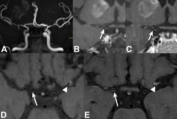
Fig 1. 51-year-old female patient with neuroborreliosis, bilateral neuritis of 3rd nerve, and right MCA territory lenticulostriate infarct.
TOF MRA (targeted MIP, A) shows bilateral medium-grade long-segmental stenosis of intradural ICAs and left MCA M1 segment, and high-grade right MCA stenosis. 2D and 3D VWI in coronal and axial views: Thin concentric VWE (white arrows) of right supraclinoid ICA is barely visible on 3D (B, D) and clearly visible on 2D (C, E) VWI. Concentric VWE at left MCA bifurcation/proximal M2 (white arrowheads) is well visualized on both 2D (E) and 3D VWI (D).

