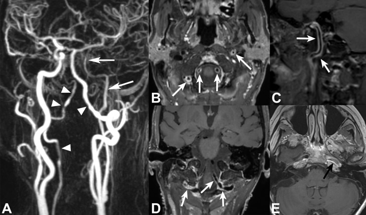
Fig 2. 68-year female patient with giant cell arteriitis and left MCA territory infarcts.
CE-MRA of supraaortic arteries (A) shows bilateral long segmental irregular VA stenosis involving right V3 and V4 as well as left V4 segments (white arrowheads) and long-segmental stenosis of left extra- and intracranial ICA (white arrows). 3D VWI with multiplanar views (B-D) depicts multiple areas of long-segmental concentric VWE in bilateral V3 and V4 segments and left cervical/petrosal ICA (white arrows). Due to limited coverage, 2D VWI shows only parts of the enhancing lesions in left petrosal ICA segment (black arrow in E).

