
Figure 1.
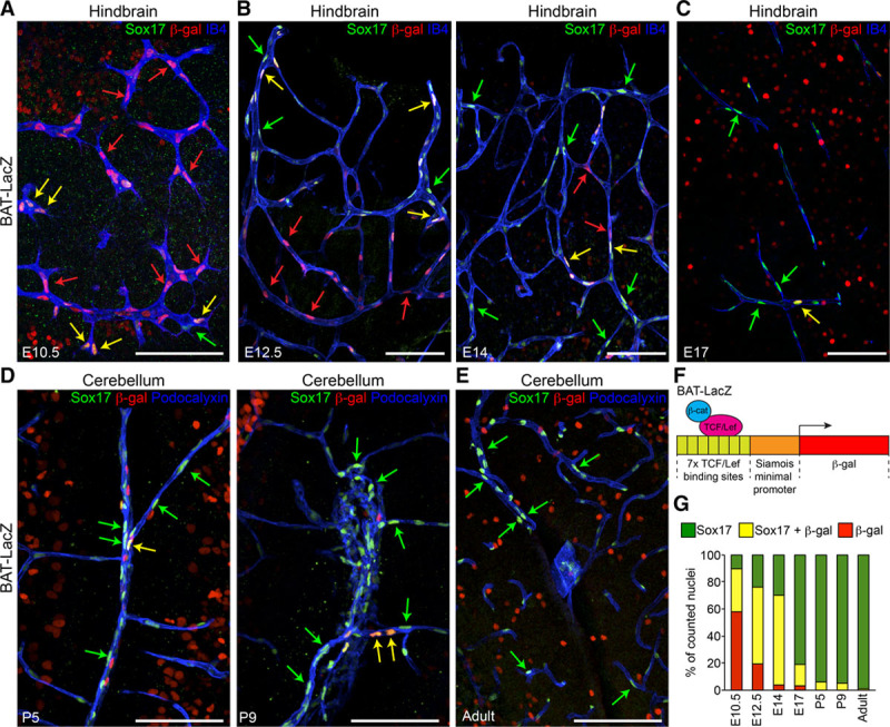
Canonical Wnt/β-cat (β-catenin) signaling and Sox17 (SRY [sex-determining region Y]-box 17) expression show distinct time courses in endothelial cells (ECs) during brain angiogenesis and vessel maturation. A–E, Confocal analysis of the brain microvasculature of BAT (β-catenin-activated transgene)-LacZ reporter embryos, pups, and adult mice. Whole-mount (A and B), vibratome sections (C), and cryosections (D and E) were stained for β-gal (β-galactosidase, red), to detect β-cat–induced expression of the Lac-Z reporter construct, Sox17 (green), and IB4 (isolectin B4, blue, A–C) or Podocalyxin (blue, D–E). Green, red, and yellow arrows indicate Sox17 staining, β-gal–positive nuclei, and double positive nuclei, respectively. Positive nuclei to β-gal outside the vascular system indicate active Wnt signaling in the brain parenchyma. Scale bar: 100 µm. F, Schematic representation of the BAT-LacZ reporter construct (see Methods). G, Quantification of nuclei positive for Sox17 only (green), β-gal only (red), or double-labeled (yellow; n=3–5 animals for each time point). These indicate different patterns of expression between Sox17 and β-cat signaling. LEF/TCF indicates lymphoid enhancer factor/T-cell factor.