
Figure 8.
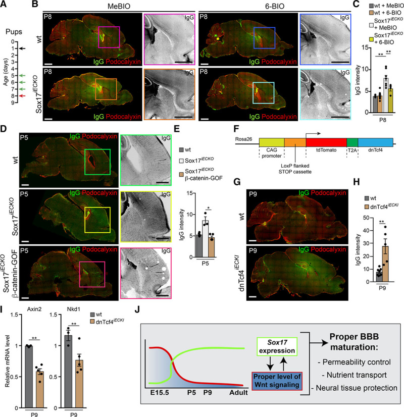
Stabilization of β-cat (β-catenin) signaling restores vascular defects induced by Sox17 (SRY [sex-determining region Y]-box 17) inactivation. A, Scheme of tamoxifen injections in the pups at P1 (black arrow), (2′Z,3′E)-6-bromoindirubin-3′-acetoxime (6-BIO; or 1-methyl- bromoindirubin [MeBIO]) injections at P5-7 (green arrows), and analysis of brain P8 (red arrow). B, Confocal images of sections stained with Podocalyxin (red) and IgG (green or black in magnification) of wild-type (wt) and Sox17iECKO pups revealed a complete rescue of IgG leakage in mutant brains treated with 6-BIO. Scale bar: 1 mm. C, Quantification of IgG leakage in brain as in B (n=3 for each experimental condition, mean±SEM, **P<0.01 ANOVA and Tukey post hoc analysis). D, β-cat stabilization rescue Sox17iECKO blood-brain barrier (BBB) defect. Confocal images of section stained with Podocalyxin (red) and IgG (green) from wt, Sox17iECKO, and Sox17iECKO/β-cat–gain of function (GOF) brains from P5 pups. The presence of IgG leakage (black [magnified]) reveals high permeability in Sox17iECKO brains. In the double mutant Sox17iECKO/β-cat GOF pups, IgG leakage is reduced to control levels. Scale bar 1 mm. E, Quantification of IgG extravasation indicates more prominent leakage in Sox17iECKO brains (n=6 wt and 3 Sox17iECKO and 5 Sox17iECKO/β-cat-GOF, mean±SEM, *P<0.05, Mann-Whitney test). F, Schematic representation of the td-Tomato-T2A–dominant-negative Tcf4 (transcription factor 7 like 2, T cell-specific, HMG box; dnTcf4) construct. See Methods for details (CAG, CMV immediate enhancer/β-actin). G, Confocal images of brain microvasculature. IgG (green) and Podocalyxin (red) staining of sections of wt and inducible endothelial-specific dominant-negative Tcf4 (dnTcf4iECKI) P9 pups. The presence of IgG reveals higher permeability in dnTcf4iECKI. Scale bar 1 mm. H, Quantification of IgG leakage indicates more prominent leakage in dnTcf4iECKI brains (n=8 wt and 5 dnTcf4iECKI, mean±SEM, **P<0.01, Mann-Whitney test). I, Real-time quantitative polymerase chain reaction analysis of 2 Wnt/β-cat target genes (Axin2 and Nkd1) expressed in brain endothelial cells isolated from dnTcf4iECKI P9 pups (n=3 wt and 4 dnTcf4iECKI, mean±SD **P<0.01, 2-tailed t test). J, Schematic model of the cross-talk between Wnt/β-cat and Sox17 signaling. Wnt signaling declines after birth and triggers activation of Sox17 that in turn control brain vascular permeability. A calibrated cross-talk of these 2 transcription pathways is necessary to prevent uncontrolled effect and pathogenic conditions.