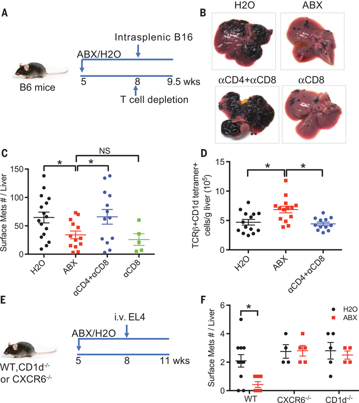
Fig. 3. Hepatic NKT cells mediate the inhibition of liver metastasis in mice.
(A to D) B16 tumor cells were intrasplenically injected into mice pretreated with ABX or H2O. One day before tumor injection, mice were given intraperitoneal injection of a combination of antibodies to CD4 (anti-CD4) (500 μg per mouse) and anti-CD8 (200 mg per mouse) or anti-CD8 alone (200 μg per mouse). Liver surface metastatic nodules were counted. Representative images are shown. Data represent mean ± SEM of two pooled experiments. n = 16 for H2O, 14 for ABX, 13 for anti-CD4 + anti-CD8, 5 for anti-CD8. P < 0.05, one-way ANOVA. (E and F) Loss of hepatic NKT abrogated the inhibition of liver metastasis caused by ABX. CXCR6−/−, CD1d−/−, or wild-type mice pretreated with ABX or H2O were given EL4 tumor cell tail vein injections. Liver surface metastatic nodules were counted. Data represent mean ± SEM of two pooled experiments. n = 10 for Wt-H2O, 7 for Wt-ABX, 4 for CXCR6−/−-H2O, 5 for CXCR6−/−-ABX, 5 for CD1d−/−-H2O, 4 for CD1d−/−-ABX. P < 0.05, two-way ANOVA.

