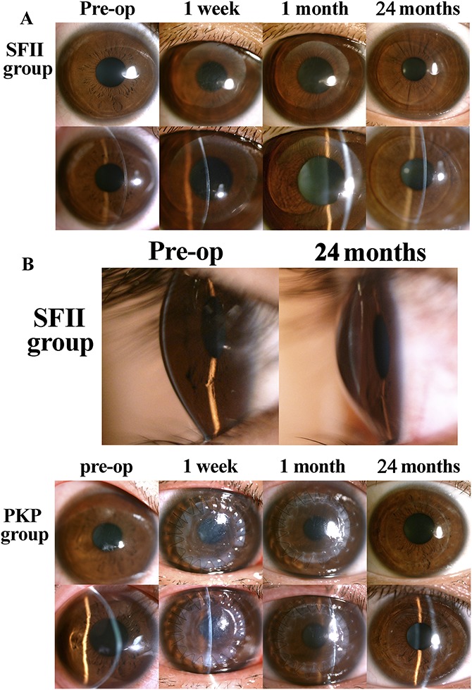
FIGURE 2.

Representative appearance of the eye on slit-lamp microscopy in both groups. A, The surrounding area of the host corneal tissue maintained its transparency in all recipient eyes. At 1 week after surgery, there was pronounced stromal edema around the implant. Graft edema greatly subsided after 1 month, and the implant boundary was smooth and visible. B, By 24 months after surgery, total integration of the implant and the host stroma was evident in all recipients undergoing the SFII procedure, and there was no visible tissue boundary. C, Graft edema and ciliary redness at 1 week postoperatively. The epithelium regenerated within 1 month, but the margin of the graft still exhibited edema. At 24 months postoperatively, graft edema subsided with a smooth graft–host junction.
