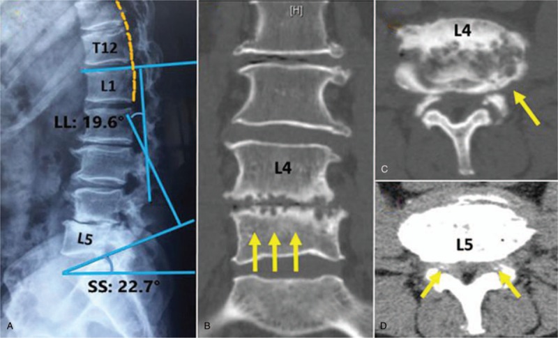
Figure 1.

(A) X-ray shows the narrowed interspace between L4-5, retrolisthesis at L4, and kyphosis in thoraco-lumbar spine. (B) Sagittal CT illustrates subchondral bones of L4 lower endplate and L5 upper endplate appearing as punched-out liked bone destruction with peripheral calcification. (C–D) Phase axial CT shows irregular erosion in vertebral body and lower endplate of L4, swelled articular soft tissue, and punctuate high density in bilateral yellow ligament. CT = computed tomography.
