
Figure 2.
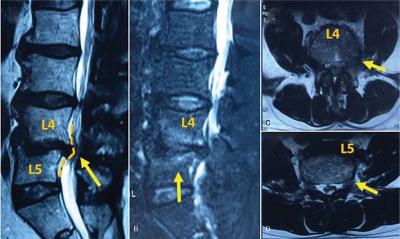
(A) MRI demonstrates the dural sac is compressed obviously. (B) Fat-suppressed T2-weighted image shows the lesion area surrounded by high signal. (C–D) Phase axial MRI illustrates the structural changes and inflammatory responses at L4-5 intervertebral disc. MRI = magnetic resonance imaging.