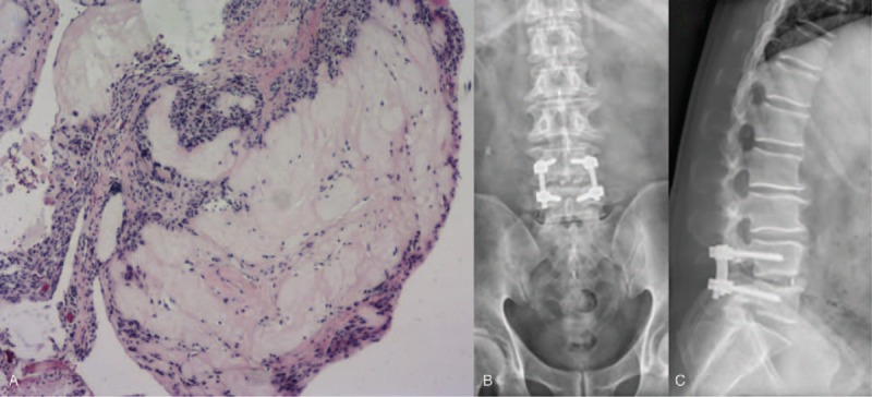
Figure 3.

(A) Intraoperative pathology (L4-5 disc): fibroblasts, inflammatory granuloma composed of lymphocytes, and foreign body giant cells are surrounded by a few urate crystals, which suggesting tophaceous gout ((hematoxylin-eosin, × 100). (B–C) Postoperative X-ray shows the location of the internal fixation and interbody fusion cage is satisfied and the sequence of spine is well reconstructed.
