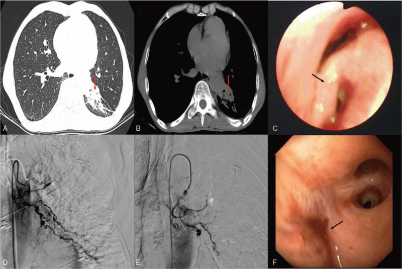
Figure 1.

(A and B) Chest computed tomography showing bronchial stenosis in the left lower lobe, accompanied with local atelectasis (red arrow), emphysema, pulmonary bullae, and local thickened pleura. (C) Bronchoscopy showing a slit-like stenosis of the dorsal segment of left lower lobe, edematous, smooth mucosa, and widening of carina (black arrow), no abnormal vessels or active bleeding is seen. (D) Selective bronchial arteriogram showing a dilated, tortuous left lower lobe bronchial artery and profusely hypervascularized dorsal segment of left lower lobe. (E) Transcatheter embolization of the hypertrophic bronchial artery using poly-vinyl alcohol particles (PVA) 500 μm in diameter. After embolization, DSA reveals complete disappearance of the abnormal artery. (F) The opening of left lower bronchial lobe is occluded, but there is no active hemorrhage after BAE (black arrow).
