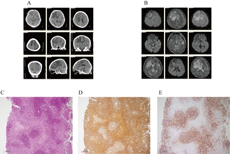
Fig. 3.
A and B. CT for case 11 demonstrated a 3-cm, low-density area with a ring enhancement lesion in the right anterior lobe, and multiple low-density areas in the right occipital, left anterior, and temporary lobes (A). Furthermore, MRI revealed a 3-cm, heterogeneous high-intensity area, and multiple T2 high-intensity lesions in the right occipital, left anterior, and temporary lobes (B). These observations suggested multiple brain metastases or PTLD in the brain.
C, D and E. On histology the brain biopsy specimen of case 11, the abnormal lymphoid cells had larger and hyperchromatic nuclei, and had diffusely proliferated predominantly in the perivascular region with marked necrosis (C). Immunohistochemically, the cells were positive for L26 (CD20), CD79a, MUM1, and Bcl-2, and negative for CD3, UCHL1 (CD45Ro), CD5, CD10, and Bcl-6, indicating non-GC type diffuse large B-cell lymphoma (DLBCL) (D). Moreover, all lymphoid cells expressed EBV markers, as assessed by EBER-FISH (E). Thus, these findings are consistent with monomorphic post-transplant lymphoproliferative disorders [DLBCL with EBV (+)].

