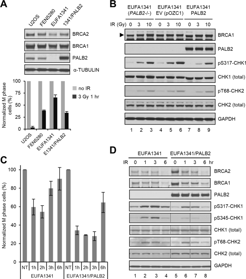
Figure 3.

G2/M checkpoint defect of PALB2-deficient human fibroblasts and its rescue by re-expression of wt PALB2. (A) G2/M checkpoint activation in U2OS, FEN5280, EUFA1341, and EUFA1341 cells reconstituted with wt PALB2. Upper panel, representative western blots showing expression levels of PALB2, BRCA1 and BRCA2 in the cells; lower panel, relative mitotic indexes of the cells before and 1 hr after 3 Gy of IR. Data shown are means ± SEMs of the relative mitotic indexes from n>3 independent experiments. (B) Dose-dependent CHK1 and CHK2 phosphorylation in blank, empty vector-harboring and PALB2-reconstituted EUFA1341 cell lines. Cells were collected before or 1 hr after 3 or 10 Gy of IR; total and phosphorylated proteins were analyzed by western blotting. (C) Checkpoint maintenance in blank and reconstituted EUFA1341 cells. Cells were treated with 3 Gy of IR and mitotic cells were measured before and at 1, 2, 3 and 6 hr after IR. Data shown are means ± SEMs of the relative mitotic indexes from 3 independent experiments. (D) Kinetics of CHK1 and CHK2 phosphorylation in blank and reconstituted EUFA1341 cells. Cells were treated with 3 Gy of IR and collected at indicated time points; total and phosphorylated proteins were analyzed by western blotting. The lanes between dotted vertical lines were loaded in the wrong order, which were reversed back using Adobe Photoshop.
