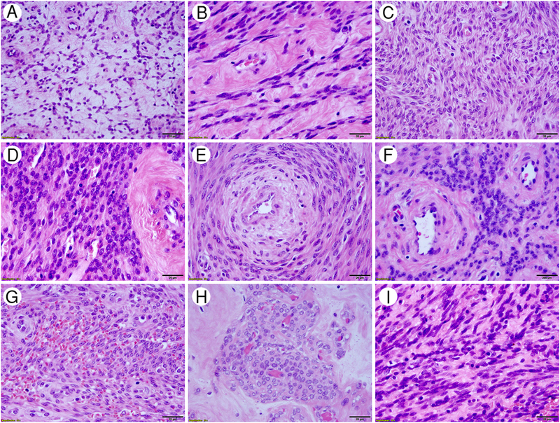
Figure 2.

Cytologic features of HLM. High-power fields in H/E stained slides revealed that HLM have tumor cells predominantly with relatively small, round or oval nuclei. Tumor cells are seen in various tumor-patterns as presented in reticulated (A), sheath-like (B), streaming (C), cellular (D), concentric (E) and perivascular (F) components; as well as with red blood cell extravasation (G), perivascular pseudonodules (H), and spindled (I) components. Magnification was indicated in right lower corner of black bar (20 μm).
