
Figure 3.
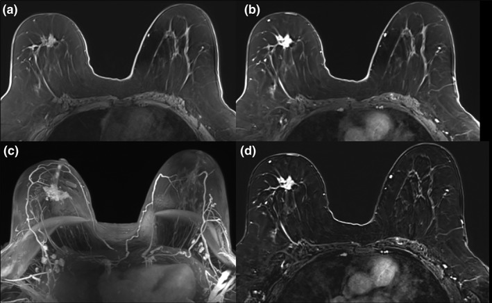
Invasive lobular carcinoma (ILC) in the right breast of a 59‐year‐old patient. The irregular, spiculated 3.5 cm mass (A) demonstrates heterogeneous contrast enhancement on early contrast‐enhanced axial T1W (B), maximum intensity projections (MIP) (C), and subtraction images axial T1W postcontrast (D) and was accurately diagnosed with an abbreviated MRI protocol.