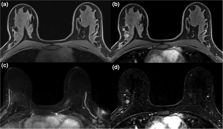
Figure 5.

Fibroadenoma in the right breast in a 26‐year‐old patient with a BRCA2 gene mutation (A). On high‐risk screening MRI there was a 0.9 cm mass lesion in the right breast, showing homogeneous contrast enhancement with a type I enhancement curve in the postcontrast axial T1W (B) and subtraction images axial T1W postcontrast (D). This benign lesion was not depicted on maximum intensity projections (MIP) images (C).
