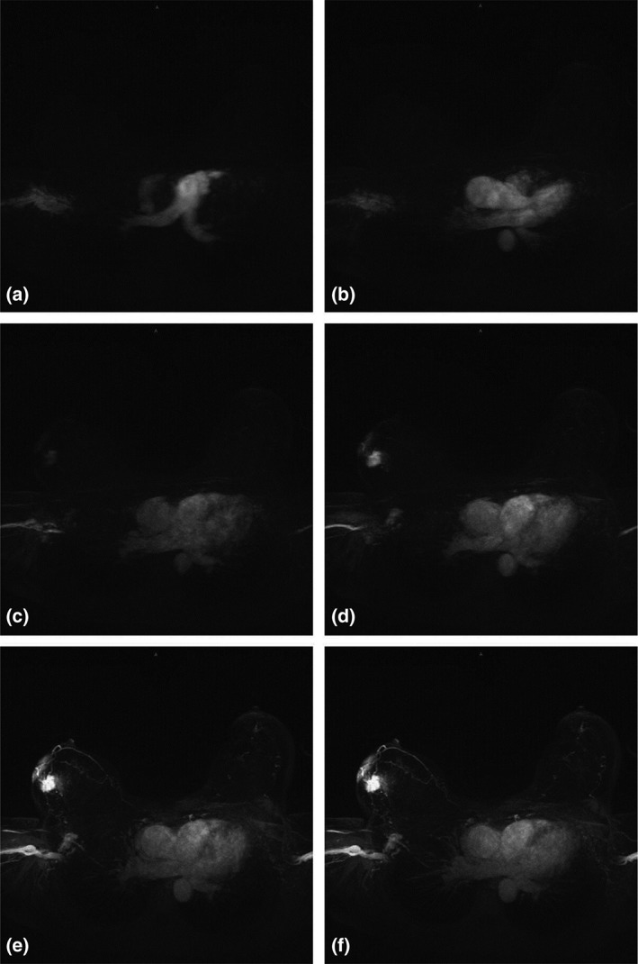
Figure 6.

Selected stills (maximum intensity projections) from a movie of contrast inflow. A: Only the pulmonary artery enhances. B: The contrast has reached the aorta. C: The tumor in the right breast starts to enhance, just as the overlying infiltrated skin. D: The tumor stands out like a light bulb in a further empty breast. E: The draining veins become visible. F: Minimal normal glandular tissue enhancement is seen. Modified with permission from: Mann RM, Mus RD, van Zelst J, et al. Invest Radiol 2014;49:579–585.
