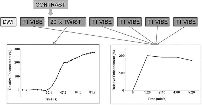
Figure 7.

Schematic drawing of the breast MRI scan protocol: The TWIST acquisitions allow evaluation of the contrast inflow in the lesion, whereas the VIBE acquisitions are used for three‐timepoint analysis, creating the classic contrast enhancement versus time curve. Modified with permission from: Mann RM, Mus RD, van Zelst J, et al. Invest Radiol 2014;49:579–585.
