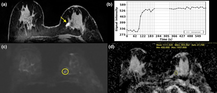
Figure 9.

Invasive ductal carcinoma (IDC) G1 in the left breast medial in a 71‐year‐old woman. (A) On DCE‐MRI there is a 10mm irregular‐shaped and marginated lesion (arrow) with (B) an initial fast/plateau enhancement (II); DCE‐MRI findings were classified as suspicious for malignancy (BI‐RADS 4). DWI was false negative as none of the readers called this lesion on DWI alone. However, when read as mpMRI combining DCE‐MRI and DWI, readers identified a (C) hyperintense correlate (circle) with (D) ADC values measuring 1.111 × 10‐3 mm2/s, which further confirmed malignancy.
