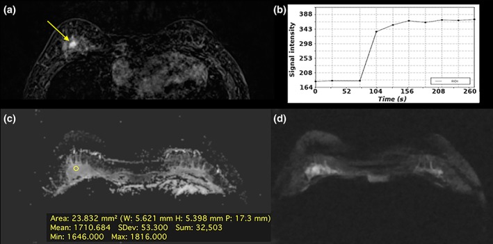
Figure 10.

Fibroadenoma in a 39‐year‐old woman, central in the right breast: (A) The irregular‐shaped and partially irregularly marginated 7 mm mass demonstrates (B) an initial medium/persistent (II) slightly homogenous contrast enhancement and was classified as suspicious (BI‐RADS 4). (C) On DWI there is no focal restricted diffusivity and (D) no decreased ADC values (1.710 × 10⁻³ mm2/s). DCE‐MRI and DWI were discordant. According to the BI‐RADS‐adapted reading algorithm, the BI‐RADS assessment category assigned based on DCE‐MRI was overruled. Multiparametric MRI correctly classified the mass as benign and would have obviated an unnecessary breast biopsy.
