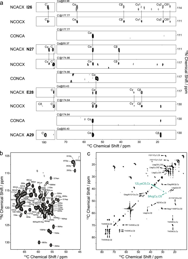
Figure 2.
Spectra used for resonance assignment of DGK by MAS NMR. (a) Representative sequential assignment of U-13C,15N-DGK from I26 to A29 based on a set of dipolar-coupling based 3D NCACX, NCOCX and CONCA spectra. Each set of three spectra stands for a Cx[i-1]-N[i]-Cx[i] spin system. For example, the N27 NCACX peaks are linked to the I26-N27 CONCA peak through the same N and Cα and the I26 NCOCX peaks are connected to the I26-N27 CONCA peak via the same N and C’, thus generating a Cx[i-1]-N[i]-Cx[i] system. It is associated with the prior system through all carbon shifts of I26 that are visible in both NCACX and NCOCX spectra. All detailed experimental NMR parameters are summarized in Suppl. Table 4. (b) 2D NCA spectrum of U-13C,15N-DGK with all assigned peaks labelled. (c) 2D scalar-coupling based 13C-13C TOBSY of U-13C,15N-DGK with tentative assignments. INEPT and TOBSY were used for 1H-13C heteronuclear polarization and 13C-13C homonuclear mixing, respectively. Peaks for Arg9 and Lys12 are highlighted green, since they could be assigned unambiguously. Amino acids that correspond to the His-tag are labelled by ‘tag’.

