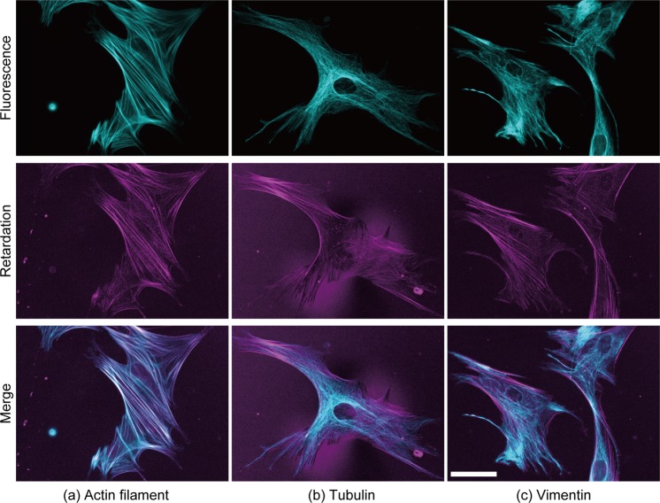
Figure 1.
Typical images of fluorescently labelled cytoskeletons and the retardation of VSMCs. Three major cytoskeletal components, i.e., (a) actin, (b) β-tubulin, and (c) vimentin, were stained with fluorescent dyes in VSMCs. The fluorescence images shown in cyan (top row) and the retardation images shown in magenta (middle row) are merged in the bottom row. White colours in the merged images show the co-localization between the cytoskeleton and retardation. Bar in the bottom right panel = 50 µm and applies to all images.

