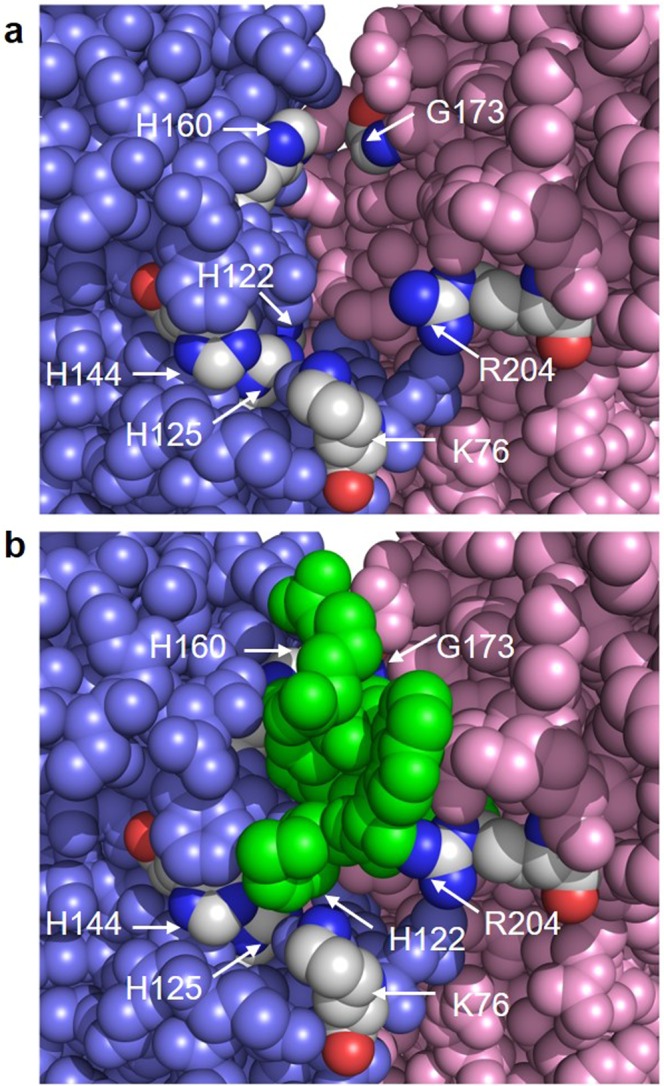
Figure 5.

Location of peptide CR20 and conserved residues implicated in catalysis. (a) View into one of the three peptide-binding cavities of the LpxA trimer with the peptide CR20 hidden. LpxA subunits are colored pink, slate, or green (not visible) as in Fig. 1. Key residues implicated in catalysis or substrate binding by mutagenesis are colored according to element with carbons in grey. (b) Same view as above but with peptide CR20 present in green. Conserved LpxA residues are labeled in white. There is some space between peptide CR20 and the conserved residues H125, H144, H122 and K76. Peptide CR20 is interacting with G173 and H160, these residues are largely hidden from view. In the complete model, a few water molecules are located in the space beneath peptide CR20 (not shown).
