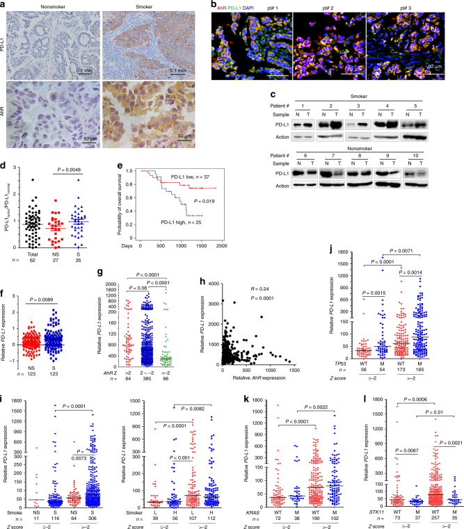
Fig. 4.
A threshold level of AhR is critical to increased PD-L1 expression. a The expression of PD-L1 and AhR was detected by IHC assay in NSCLCs. b Immunofluorescence assays of smoker patients’ formalin-fixed paraffin embedded (FFPE) 5-μm sections using antibodies against PD-L1 and AhR, and DAPI. Arrow, cancer cells; *, non-cancerous cells. c Western blot analyses of lysates of tumor (T) and adjacent normal (N) lung tissues harvested from NSCLCs (n = 62). d Quantification of the ratios of PD-L1 in tumors to PD-L1 in counterpart normal lung tissues in smoker and nonsmoker patients. Determined by densitometry analyses of immunoblot bands in (c). NS, nonsmoker; S, smoker. P value, Student’s t test. e Overall survival of the 62 patients. P value, log-rank test. f PD-L1 expression in an Oncomine report. g The expression of PD-L1 and AhR in DNA microarray data of TCGA datasets. AhR z score was calculated as: (RNA-Seq by Expectation Maximization (RESM) in tumor - mean of RESM values in normal)/Standard deviation of RESM values in normal). P value, Mann-Whitney test. h The association between PD-L1 and AhR in patients. Data are from TCGA datasets. i The smoking status, PD-L1 expression, and AhR Z score in NSCLCs. j–l The expression of PD-L1, AhR Z score, and TP53 (j), KRAS (k), or STK11 (l). P values in i-l, Mann-Whitney test. WT wild type, M mutant, H heavy smoker, L light smoker

