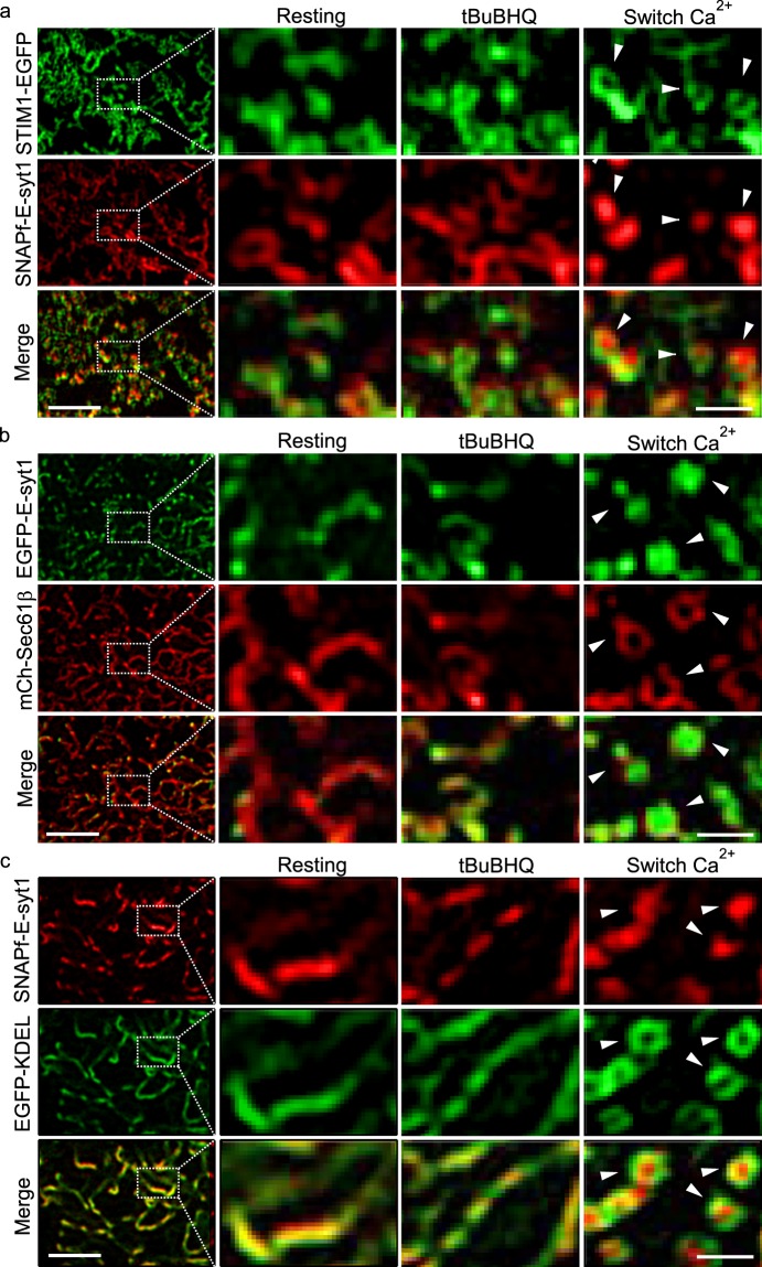
Figure 3.
Ca2+ influx via SOCE activates E-syt1 to reshape punctate ER-PM MCSs into ring-shaped structures under TIRF-SIM. (a–c) Representative TIRF-SIM images of cortical ER in HEK293 cells co-transfected with SNAPf-E-syt1 and STIM1-EGFP (a), EGFP-E-syt1 and mCherry-Sec 61β (b), or SNAPf-E-syt1 and EGFP-KDEL (c). The left panels show images of cells under resting conditions at a lower magnification; boxed areas are magnified and shown on the right. From left to right are cells under resting conditions, store depletion and store replenishment. All these figures are representative of four independent experiments. Scale bars, left 2 μm; right 0.5 μm. Related videos were also shown as Supplementary Videos S2–S4.

