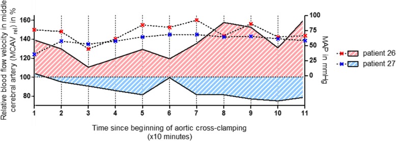
Fig. 2.
Individual relative blood flow velocities in middle cerebral artery during cardiopulmonary bypass. Blood flow velocity in the right middle cerebral artery (MCAV) was measured using transcranial Doppler sonography. Individual values, assessed during cardiopulmonary bypass (CPB) every 10 min, were normalized to the baseline value obtained before cannulation of the aorta and was termed MCAVrel. The figure shows the relative cerebral blood flow velocities in right MCA during CPB (solid lines, plotted on left Y axis), together with the corresponding mean arterial blood pressure (MAP) values (dashed lines, plotted on right Y axis), from two representative patients over the time. MCAV of patient no. 27, which developed no delirium, was below his individual baseline value almost the whole time during CPB. By contrast, the MCAV of patient no. 26, developing POD, was above his baseline at any time point, with peak values exceeding 160%. Note that MAP was kept within a range between 50 and 90 mmHg almost all the time in both patients

