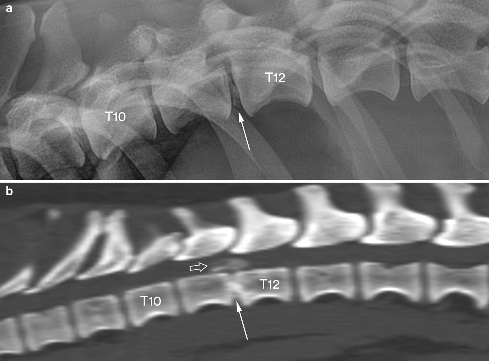
Fig. 2.

Lateral radiograph (a) and a sagittal MPR CT image in bone reconstruction algorithm (b) of the caudal thoracic and cranial lumbar vertebral column segments in dog no. 24. The radiograph shows a slight degree of calcification in the T11–T12 disc space (arrow), whereas the CT image reveals calcified material in the vertebral canal (open arrow) above a calcified T11–T12 disc space (solid arrow)
