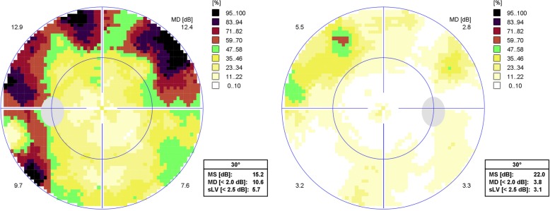
Fig. 2.
30° Octopus visual field examination showing marked superior and inferior arciform scotomata in the left eye (left image) with a mean deviation of 10.6 dB, and a mild superior arciform scotoma in the right eye (right image) with a mean deviation of 3.8 dB. (MS: Mean sensitivity; sLV: Square root of lost variance)

