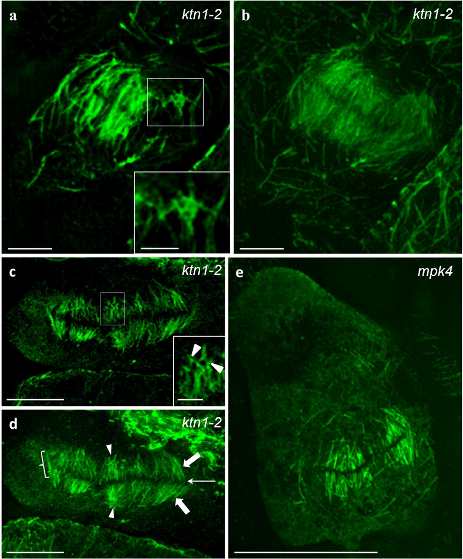
Fig. 4.

Observation of abnormal phragmoplast formation in the ktn1-2 and mpk4 mutants. a, b Single optical section (a) and maximum intensity projection (b) of an early phragmoplast of a ktn1-2 cytokinetic root cell, showing an area with extensively branched microtubules (a, inset) at the phragmoplast periphery. c, d Single optical section (c) and maximum intensity projection (d) of an aberrant late phragmoplast of the ktn1-2 mutant. Excessive microtubule branching (arrowheads) is observed within the phragmoplast (a, c; inset). e Ectopic and abortive phragmoplast of an aberrant root epidermal cell of the mpk4 mutant. Bars in e = 10 μm; c, d = 5 μm; a, b = 2 μm; insets of a and c = 1 μm
