Fig. 5.
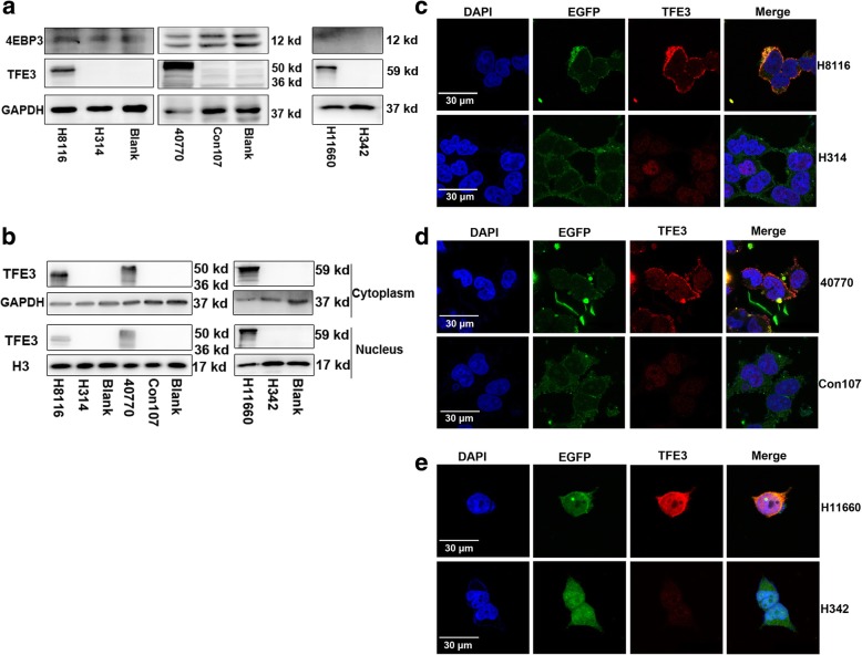
The fusion segments of TFE3 cannot be translocated to nucleus. (a) Expression levels of TFE3 and 4EBP3 were examined by immunoblot analysis. 293 T cells were transfected with plasmids H8116 (TFE3 296–575 aa), 40,770 (179–575 aa) or H11660 (full TFE3), each being compared to their relative controls by the transfection of negative control plasmids H314, Con107 and H342. (b) Expression levels of TFE3 in the cytoplasm and nucleus were examined by immunoblot analysis. Proteins expressed in the cytoplasm and nucleus were separated 48 h after transfection. GAPDH was used as a cytoplasmic internal control and H3 was shown as a nuclear internal control. (c) Cells were transfected with plasmids H8116 or H314. Expression of TFE3 (red) and EGFP (green) in 293 T cells was examined by immunofluorescence microscopy 48 h after transfection. Expression intensity of the plasmids in 293 T cells was examined based on EGFP fluorescence. Nuclei were stained with DAPI (blue). (d) Cells were transfected with plasmids 40,770 or Con107. Expression of TFE3 (red) and EGFP (green) in 293 T cells was examined by immunofluorescence microscopy 48 h after transfection. (e) Cells were transfected with plasmids H11660 or H342 for 48 h 48 h after transfection. Expression of TFE3 (red) and EGFP (green) in 293 T cells was examined by immunofluorescence microscopy. Scale bar, 30 μm

