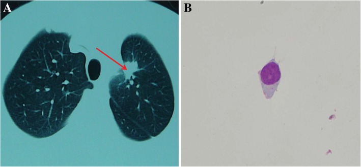
Fig. 3.
Pulmonary Magnetic Resonance imaging and CSF cytology exams, (Magnification: 1000X) after adjusting the medication based on the NGS. a Lesion in the superior lobe of the left lung was significantly reduced after adjusting the targeted drug therapy. The red arrow identifies the placeholder. Figure 2 CSF cytology exams. Magnification: 1000X. b Cell types in CSF. CSF showed 17 lymphocytes, 3 monocytes, 2 activated monocytes and 2 neutrophilic granulocytes

