Fig. 3.
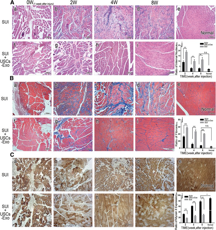
USCs-Exo promoted the recovery of the pubococcygeus muscle. A HE staining of the pubococcygeus muscle. In the SUI group, the newborn muscle fibers varied in size and shape and remained disorder (A-b, A-c, A-d). However, the muscle bundle structures in the SUI + USCs-Exo group (A-g, A-h, A-i) recovered to almost normal (A-e). Nuclear ingression was observed in both groups (A-j). B Masson staining of the pubococcygeus muscle: 8 weeks after the injection, fibrosis and muscle morphology were close to normal in the SUI + USCs-Exo group, while muscle atrophy and vicarious hypertrophy were still observed and the muscle structure was disordered in the SUI group. Fibrosis (B-j). C Immunohistochemistry analysis of the pubococcygeus muscle. Expression of myosin II (dark stained) was different in the two groups. Expression of myosin II was more in the SUI + USCs-Exo group than in the SUI group. Also, the expression of myosin II increased, gradually (C-j) (*P < 0.05, **P < 0.01, ***P < 0.001; n.s., no significance; × 100 magnification)

