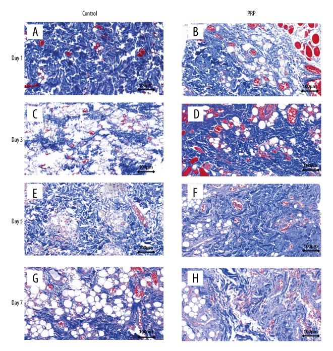
Figure 3.
Masson staining of skin flap at various time points. (A, B) No difference was found between the control group and PRP group on day 1. (C–F) Dermal collagen fibers in the control group were arranged more closely in the PRP group than in the control group. (G, H) The structure and arrangement of collagen fibers in the PRP group were better than in the control group.

