
Fig. 1.
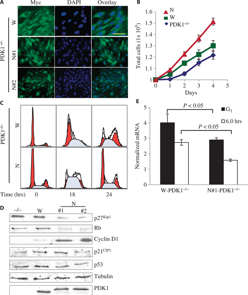
Increased proliferation and accelerated cell cycle in cells with nuclear-localized PDK1. (A) Representative confocal immunofluorescence images of three independent experiments showing the subcellular localization of myc-tagged W-PDK1−/− (W) and nuclear-localized mutant PDK1 (N) in clones of MEFs. PDK1 proteins were detected with an anti-myc antibody (green), and the nuclei were detected with 4’,6’-diamidino-2-phenylindole (DAPI; blue). Scale bar, 50 μm. (B) PDK1−/−, W-PDK1−/−, and N-PDK1−/− cells (1 × 10⁵) were seeded on 60-mm dishes, and cell numbers were counted everyday for 4 days. Values represent means ± SD (n = 3 experiments). (C) Representative flow cytometric analysis data showing the increased G1-to-S progression of N-PDK1−/− cells. MEFs of the indicated genotypes were synchronized and released from G1 phase arrest as described in Materials and Methods. Samples were collected every 6 hours, fixed, and their DNA content was measured. (D) Western blots from asynchronous MEFs of the indicated genotypes showing the amounts of important cell cycle regulatory proteins. For quantification of the three independent experiments, see fig. S3A (n = 3 experiments). (E) Graphical representation of the abundance of p27Kip1 mRNA, which was determined by quantitative polymerase chain reaction (PCR) and normalized to that of mouse TATA box-binding protein (TBP) mRNA. Values represent means ± SD (n = 3 experiments).