
Fig. 4.
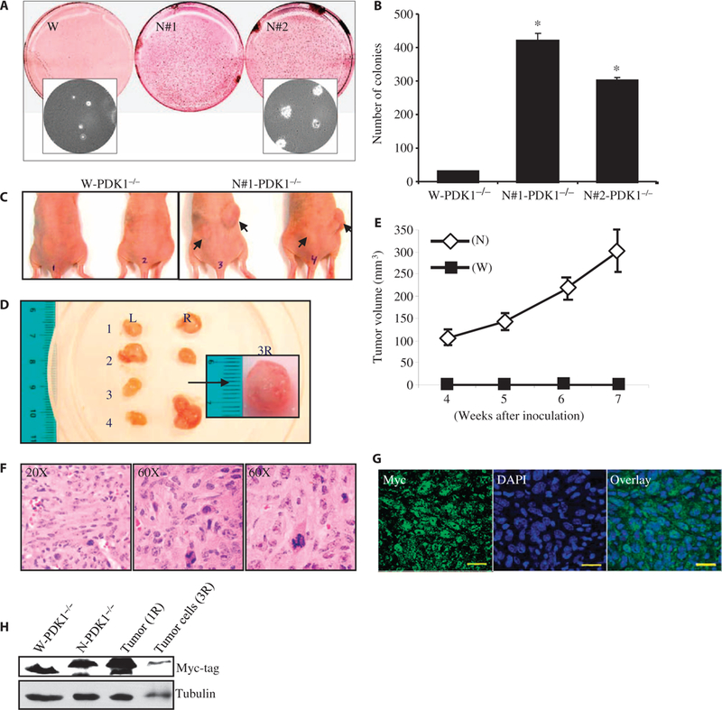
Nuclear localization of PDK1 causes oncogenic transformation of MEFs. (A) Cells (5 × 10³) were seeded on 0.4% SeaPlaque agarose plates in triplicate. Three weeks after incubation, the colonies were stained and scanned on a flatbed scanner. The inset picture was taken at ×40 magnification before the colonies were stained. (B) Quantification of soft agar colonies formed from W-PDK1−/− and N-PDK1−/− cells after 3 weeks of growth. Data are means ± SD of three independent experiments. *P < 0.005 versus W-PDK1−/−. (C) In vivo tumorigenesis by N-PDK1−/− cells. Six-week-old athymic nude mice were injected with W-PDK1−/− cells or N-PDK1−/− cells. The arrows indicate tumor growth at the injection site 7 weeks after injection. (D) Size and morphological representation of tumors isolated from mice injected with N-PDK1−/− cells. L, left flank; R, right flank. The numbers correspond to individual mice. Inset picture shows the tumor isolated from the right flank of mouse 3 (tumor 3R) for the purpose of preparing cell lines. Scale bar, 1 cm. (E) Tumor growth curve during the 7 weeks of the study. The data represent the weekly average growth of tumors measured on the right flank of five mice injected with PDK1 cells. Data are means ± SEM. (F) Representative H&E staining image of tumor 4R. (G) Immunohistochemistry (IHC) of tumor 4R with an anti-myc antibody. (H) Representative Western blotting analysis from three separate experiments in which the presence of myc-tagged nuclear PDK1 was confirmed in homogenates derived from cells (tumor ID, 3R) and tumor (tumor ID, 1R). W-PDK1−/− and N-PDK1−/− MEF lanes are controls for the tumor-derived samples.